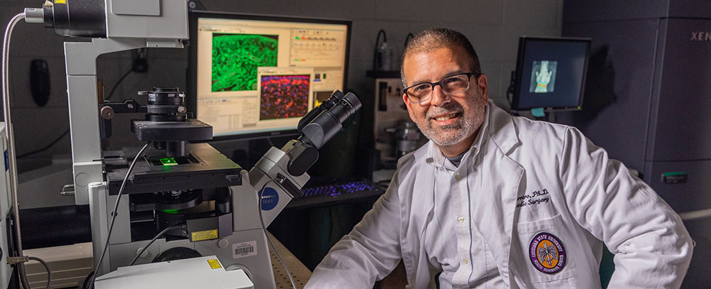
Research Team
Lab manager: TBD
Research Assistants / Undergraduate Students:
-Past: Kathryn Jordan, Sarah O'Brien, Nathaniel Beech, Jose Cruz Ayala
-Present: Christina McCarthy, Elena Plakotaris, Maria Tovar
Medical Students:
-Past: Linus Igbokwe, Jacob Davis, Jonathan Schuon, Lauren Saunee
-Present: Sydney Hodgeson, Mallory Crawford, Jose Cruz Ayala, Lauren Guillot
Headed by: Dr. Luis Marrero
Projects
First, using a platform of disparities of care in orthopedics by race, gender, and/or
age, Dr. Marrero is looking into differences in the underlying molecular mechanisms
that drive fibrosis during joint disease and following the trauma of joint replacement
surgery between local subgroups of patients affected by osteoarthritis. Specifically,
he is studying the differential modulation of pro- (e.g. TGFbeta-1, IL13) and anti-fibrotic
(e.g. IL10) factors that are known to participate in extracellular matrix remodeling
during injury and repair of the joint by means of histopathology, protein profiling,
and transcriptome analyses of normal and diseased components of joints harvested during
surgery and banked in our Musculoskeletal Specimen Repository. He then correlates
these findings with measures of joint stiffness, pain, and function gathered from
each patient, thus adding clinical significance to the project. Second, in collaboration
with Drs. Keith van Meter and Mimi Sammarco from the Division of Emergency Medicine
at UMC and the Tulane Department of Surgery, respectively, Dr. Marrero is testing
the effects of hyperbaric oxygen treatment (HBOT) in rescuing musculoskeletal components
in a pig model of traumatic limb injury and ischemia. Dr. Marrero assesses delivery,
tissue phenotype by histology and protein analyses, and novel approaches to measuring
metabolic activity (e.g. ATP biosynthesis in vivo). Third, in collaboration with
the Departments of Pathology and Pulmonary Care at the Medical University of South
Carolina, Dr. Marrero studies the effect of a novel cryopreservative of tissues for
transplantation. He analyses the integrity of vascular components harvested from donor
limbs and their viability for seeding into large allografts used in surgical correction
of massive skeletal defects.
Resources
Facilities and equipment for histopathology, cell culture, biophotonic imaging in
vivo, and xray. Microscopy systems for brightfield, epifluorescence, confocal, multiphoton,
and laser scanning microdissection. For more information visit our resources page.

