Case of the Month - April 2024
Yu Liu, MD; Liz Yang, MD; Ridin Balakrishnan, MD
A 52-year-old female with a history of abnormal uterine bleeding and an endometrial
mass detected on transvaginal ultrasound was admitted to the hospital for a hysteroscopy,
dilation and curettage, and myomectomy. Intraoperative findings of significance included
a 4.0 cm probable fibroid in the lower uterine segment. A resectoscope was used to
remove approximately 90% of this lesion. Multiple fragments of white-tan to pink-tan
fibroid-like tissue were sent to pathology for assessment. Microscopic sections demonstrated
a myometrial based neoplasm composed of spindled cells in vague fascicles surrounded
by delicate thin-walled vessels. However, areas of epithelioid morphology with clear
to eosinophilic and granular cytoplasm were also noted. Representative hematoxylin
and eosin (H&E)-stained sections at low and high power are illustrated in the Figure
1 below.
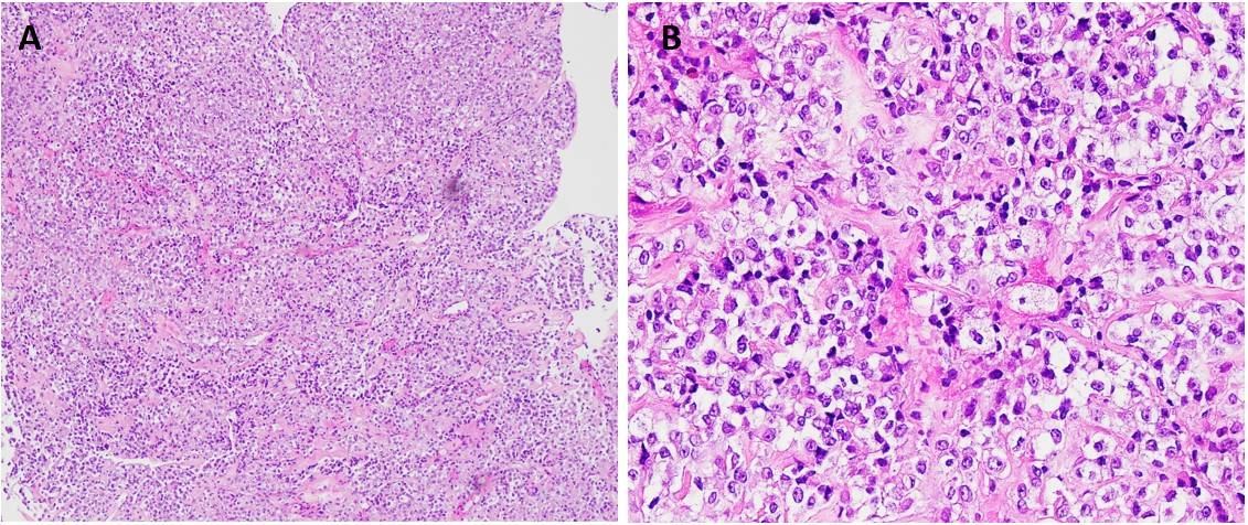
Figure 1: Epithelioid cells with clear to eosinophilic granular cytoplasm are arranged in sheets surrounded by delicate thin-walled vessels (A: H&E,100x; B: H&E, 400x)
By immunohistochemical (IHC) stains the lesional cells were patchy positive for Desmin (Figure 2A), ER/PR, p16, Calretinin, Melan A (Figure 2B) and HMB45 (Figure 2C), with rare expression of CK7. No significant expression of pan-cytokeratin, EMA, PAX8, p63, p40, GATA-3, S100, SOX10, CD117, and DOG1 was seen. P53 demonstrated wild-type staining. Some IHC stains are illustrated in the Figure 2 below.
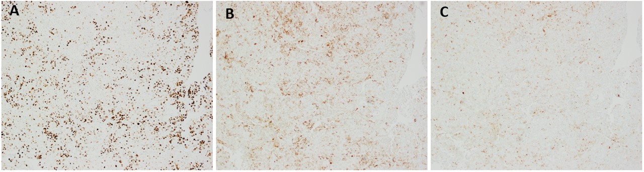
Figure 2: IHC stains (100x): (A) Desmin; (B) Melan A; (C): HMB45
Following the diagnosis, the patient returned to the hospital for a total laparoscopic hysterectomy and bilateral salpingo-oophorectomy. Gross examination of the resected specimen revealed a pink-tan, pedunculated, friable soft mass measuring 3.5 x 3.0 x 2.7 cm, attached to both the anterior and posterior left endometrium, occupying the entire endometrial canal. Representative H&E-stained sections from the mass are illustrated in Figure 3 (A, B: 200x) below. Most of the tumor in the resection resembled the spindled morphology of the lesion in the biopsy. Additionally, tumor cells focally demonstrated a perivascular radial distribution.
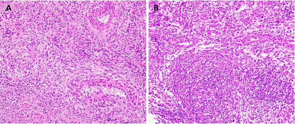
Figure 3: Tumor cells exhibit a perivascular distribution, with certain areas displaying spindled cells with eosinophilic cytoplasm organized into short fascicles (A, B: H&E, 200x)
The mass had an immunoprofile similar to the prior biopsy: SMA (patchy), Caldesmon (focal), Desmin (focal), HMB45 (diffuse), ER (focal), PR (patchy), and CD10 (focal). Diffuse expression of Cathepsin K (Figure 4A) was noted. Additional IHC performed for TFE3 (Figure 4B) was negative.
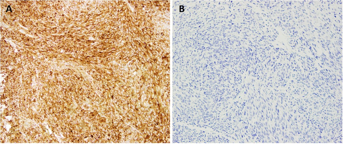
Figure 4: IHC stains (200x): (A) Cathepsin K; (B) TFE3.
What is the most likely diagnosis?
1. Epithelioid smooth muscle tumor
2. Alveolar soft part sarcoma
3. Perivascular epithelioid cell tumor
4. Malignant melanoma
5. Poorly Differentiated Endometrial Carcinoma