Publications and Center Leadership Highlighted on Journal/Magazine Covers
CNS Neurosci 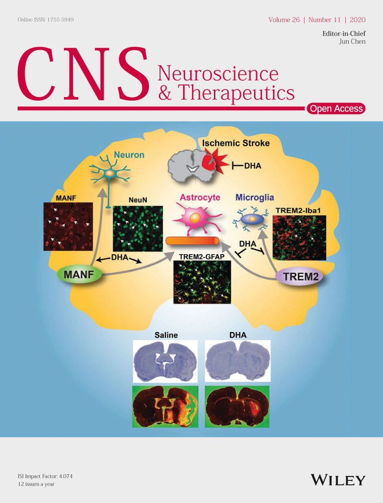 Ther. 2020;26:1155-1167.
DHA modulates MANF and TREM2 abundance, enhances neurogenesis, reduces infarct size,
and improves neurological function after experimental ischemic stroke
Ludmila Belayev, Sung-Ha Hong, Raul S. Freitas, Hemant Menghani, Shawn J. Marcell,
Larissa Khoutorova, Pranab K. Mukherjee, Madigan M. Reid, Reinaldo B. Oria, Nicolas
G. Bazan
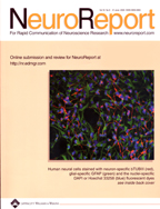
COVER: image is based on the Original Article “DHA modulates MANF and TREM2 abundance, enhances
neurogenesis, reduces infarct size, and improves neurological function after experimental
ischemic stroke” by Ludmila Belayev et al., https://doi.org/10.1111/cns.13444.

Matters o f the Mind A New Finding and a New Hope Alzheimer's
Nicolas G. Bazan
 Journal Lipid Research , 2020;61:10,
Docosanoid signaling modulates corneal nerve regeneration: effect on tear secretion,
wound healing, and neuropathic pain
Thang L. Pham and Haydee E.P. Bazan
COVER: The cornea is densely innervated, mainly by sensory nerves of the ophthalmic branch
of the trigeminal ganglion (TG). These nerves play a pivotal role in corneal health
and in the pathological genesis of diseases such as neurotrophic keratitis, corneal
ulceration, dry eye disease, and neuropathic pain. Using animal models of corneal
nerve damage, treatment with pigment epithelium-derived factor (PEDF) plus docosahexaenoic
acid (DHA) stimulates nerve regeneration through a mechanism that involves the synthesis
of docosanoids including neuroprotectin D1 (NPD1) and the new resolvin stereoisomer
RvD6i. These lipid mediators activate the corneal-TG axis, resulting in an increase
of corneal wound healing, sensation recovery, and tear secretion without any exacerbation
of corneal pain. The actions of these docosanoids open avenues of therapeutic exploration
to re-establish corneal homeostasis in many clinical pathologies. (See Pham and Bazan.)
Invest Ophthalmol Vis Sci. 2019;60:2449-2460.
Remodeling of Substance P Sensory Nerves and Transient Receptor Potential Melastatin
8 (TRPM8) Cold Receptors After Corneal Experimental Surgery
Jiucheng He, Thang Luong Pham, Azucena H. Kakazu, and Haydee E. P. Bazan
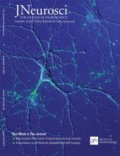
The Journal of Neuroscience , September 19, 2018 • 38(38):8110-8127
Acetylcholine, via SK Channels, Regulates CA3-CA1 Coupling
Crescent L. Combe, Carmen C. Canavier, and Sonia Gasparini
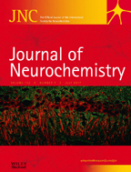 JOURNAL OF NEUROCHEMISTRY 2017;142:29-40
Functional identification of activity‐regulated, high‐affinity glutamine transport
in hippocampal neurons inhibited by riluzole
Jeffrey Erickson
COVER: Sodium-coupled neutral amino acid transporters SNAT1 and SNAT2 are considered to be the neuronal system A transporters that supply glutamine to neurons for synaptic transmission. SNAT1 is enriched in all parvalbumin-containing inhibitory neurons in the cortex and hippocampus, while SNAT2 is predominant in all pyramidal excitatory neurons. However, SNAT1 and SNAT2 are confined to the cell soma and proximal dendritic region and both are excluded from axon terminals suggesting that other unidentified neuronal glutamine transporters expressed in synapses support GABA and glutamatergic transmission. Shown is a merged confocal projection image (400X) of immunohistochemical staining in the rat hippocampal CA1 region for SNAT2 (red) with VGLUT1 (green). Non-biased imaging was provided by the LSUHSC Morphology and Imaging Core.
Read the full article ‘Functional identification of activity-regulated, high-affinity glutamine transport in hippocampal neurons inhibited by riluzole' by J. D. Erickson (J. Neurochem. 2017, vol. 142 (1), pp. 29-40) on doi: 10.1111/jnc.14046
 Research Features December 2016 Issue 103:6-13;
A Marvellous Mind
Nicolas G. Bazan
December 2016 Issue 106:74-77
“Connections: the importance of mentoring and collaboration to research success”
Nicolas G. Bazan
Journal of Lipid Research Vol 55, 2014
Spatial organization of lipids in the human retina and optic nerve by MALDI imaging
mass spectrometry
Karin A. Zemski Berry,* William C. Gordon,† Robert C. Murphy,* and Nicolas G. Bazan1,†
Department of Pharmacology,* University of Colorado Denver, Aurora, CO 80045; and
Neuroscience Center of Excellence,†Louisiana State University Health Sciences Center, New Orleans, LA 70112
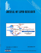
COVER: MALDI MS imaging of the macular region of the human retina. Merged MALDI positive
ion images from a cryo-section of a 47-year-old donor retina are shown at the left.
These images show unique lipid layering of phosphatidylcholine (PC) molecules: m/z
986.6 PC(50a:11)+Na, green; m/z 958.6 PC(48a:11)+Na, blue; and m/z 1070.6
PC(56:11)+Na, red. These are PCs composed of a long chain- and a very long-chain
polyunsaturated fatty acid. Docosahexaenoic acid (DHA; 22:6) is located at the sn-2
position of retina PC molecules. The red label corresponds to the photoreceptor layer,
the green label to the inner retina. Negative ion lipids are shown at top right (red
is photoreceptor layer, green is inner retina). Very long chain polyunsaturated fatty
acid-containing PCs from this same section are shown at middle right. Image at bottom
right illustrates lipid stratification with a ceramide in the retinal pigment epithelium.
(See Zemski Berry et al., p. 504.)
Invest Ophthalmol Vis Sci. 49:7, July 2008.
Association of Protein Tyrosine Phosphatases (PTPs)-1B with c-Met Receptor and Modulation
of Corneal Epithelial Wound Healing
Azucena Kakazu, Guru Sharma, and Haydee E. P. Bazan
COVER: Colocalization of the hepatocyte growth factor receptor (c-Met) and protein tyrosine phosphatase 1B (PTP1B) in human corneal epithelial cells. The cells were fixed with paraformaldehyde and immunostaining with PTP1B (green) and c-Met (red) . Hoechst was used for nuclear counterstained. The colocalization seems to be important for silencing c-Met signaling inside the cells. Azucena Kakazu, Guru Sharma and Haydee E.P. Bazan

Recent Advances in Retinal Degeneration
Book dedicated to Nicolas Bazan, 2008
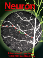
Neuron 50:291-307, April 20, 2006 DOI 10.1016/j.neuron.2006.03.016
Integrative Properties of Radial Oblique Dendrites in Hippocampal CA1 Pyramidal Neurons
Attila Losonczy and Jeffrey C. Magee
The Journal of Neuroscience , 2005 • 25(31):7121-7133 • 7121
Homeostatic Scaling of Vesicular Glutamate and GABA Transporter Expression in Rat
Neocortical Circuits
Stéphanie De Gois,1 Martin K.-H. Schafer,2 Norah Defamie1, Chu Chen,1 Anthony Ricci,1 Eberhard Weihe,2 Hélène Varoqui,1 and Jeffrey D. Erickson1
1Neuroscience Center of Excellence, Louisiana State University Health Sciences Center,
New Orleans, Louisiana 70112, and 2Department of Molecular Neuroscience, Institute of Anatomy and Cell Biology, Philipps
University Marburg, 35033 Marburg, Germany
COVER: Triple immunofluorescent staining reveals homeostatic scaling of vesicular glutamate transporters at excitatory synapses. Hyperexcitation in neocortical neuronal networks caused downregulation of VGLUT1 (red) at most synaptic terminals that face postsynaptic density 95 (blue), whereas VGLUT2 increased in some VGLUT1-containing synapses (yellow/blue) and at other sites (green/blue, green only). VGLUT2 is coexpressed with VGLUT1 in a novel class of excitatory neurons in the cerebral cortex. Their differential synaptic regulation may serve as negative or positive feedback regulators for neocortical excitatory transmission. For details, see the article by De Gois et al. in the August 3, 2005 issue (pages 7121-7133).
NEUROREPORT 16:9:21, 2005
24S-hydroxycholesterol induces inflammatory gene expression in primary human neural
cells
Piotr Alexandrov,1 Jian-Guo Cui,2 Yuhai Zhao2 and Walter J. Lukiw2,CA
1Russian Academy of Medical Sciences,Moscow113152, Russia; 2Neuroscience Center, Louisiana State University Health Science Center, New Orleans
LA 70112,USA.
The Journal of Neuroscience , 2004 • 24(49):i • i
On the Initiation and Propagation of Dendritic Spikes in CA1 Pyramidal Neurons
Sonia Gasparini,1 Michele Migliore,2,3 and Jeffrey C. Magee1
1Neuroscience Center, Louisiana State University Health Science Center, New Orleans,
Louisiana 70115, and 2Department of Neurobiology, Yale University School of Medicine, New Haven, Connecticut
06520, and 3Institute of Biophysics, National Research Council, 90146 Palermo, Italy
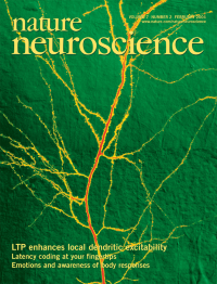
Nature Neuroscience 7:126-135 (2004)
LTP is accompanied by an enhanced local excitability of pyramidal neuron dendrites
Andreas Frick1,2, Jeffrey Magee2,3 & Daniel Johnston1,2
1Division of Neuroscience, Baylor College of Medicine, One Baylor Plaza, Houston, Texas
77030, USA. 2Marine Biological Laboratory, Woods Hole, Massachusetts 02543, USA. 3Neuroscience Center, LSU Health Science Center, 2020 Gravier Street, New Orleans,
Louisiana 70112, USA.
The Journal of Lipid Research , 2003, 44:2221
Synaptic lipid signaling: significance of polyunsaturated fatty acids and platelet-activating
factor
Nicolas G. Bazan
COVER: Simplified cartoon of phospholipases A2 at the synapse. Depolarization triggers release of glutamate, which in turn binds to the N-methyl-D-aspartate receptor and increases calcium influx into the postsynaptic neuron. Phospholipases A2 release arachidonic acid (AA), docosahexaenoic acid (DHA), and lyso-platelet-activating factor (lyso-PAF), the precursor of platelet-activating factor. sPLA2, secretory phospholipase A2. (See Bazan, p. 2221.)

The Journal of Neuroscience , 2002, 22(1):142-155
Identification of the Differentiation-Associated Na_/PI Transporter as a Novel Vesicular
Glutamate Transporter Expressed in a Distinct Set of Glutamatergic Synapses
Hélène Varoqui,1,2 Martin K.-H. Schäfer,4 Heming Zhu,1Eberhard Weihe,4 and Jeffrey D. Erickson1,3
1Neuroscience Center and Departments of 2Opthalmology and 3Pharmacology, Louisiana State University Health Sciences Center, New
Orleans, Louisiana 70112, and 4Department of Molecular Neuroscience, Institute of Anatomy and Cell Biology, Philipps
University
Marburg, D-35033 Marburg, Germany.
Aluminium and Alzheimer's Disease 2001:147-168
The Science that Describes the Link
Chapter 7 - Aluminum and Gene Transcription in the Mammalian Central Nervous System
— Implications for Alzheimer's Disease
Walter J.Lukiw
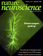
Nature Neuroscience :
Somatic EPSP amplitude is independent of synapse location in hippocampal pyramidal
neurons
Jeffrey C. Magee1 and Erik P. Cook2
1 Neuroscience Center, Louisiana State University Medical Center, 2020 Gravier St.,
New Orleans, Louisiana 70112, USA
2 Howard Hughes Medical Institute, Baylor College of Medicine, One Baylor Plaza, Houston,
Texas 77050, USA.
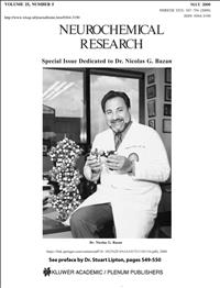
Neurochemical Research , Vol. 25, No. 5, 2000, pp. 547
Special Issue Dedicated to Dr. Nicolas G. Bazan
Molecutar Neurobiology. ISSNO893-76.4g/97/15(2!: 165-192; 1997
Vesicular Neurotransmitter Transporters: Potential Sites for the Regulation of Synaptic
Function
Hélène Varoqui and Jeffrey D. Erickson
Neurotoxicology Summer 1987; 8(2):291-301 (Cover Story)
Alterations in human linker histone-DNA binding in the presence of aluminum salts
in vitro and in Alzheimer's disease
Walter J Lukiw, Theodore PA Kruck, Donald R. McLachlan
Department of Medical Biophysics and Department of Physiology, University Of Toronto
Toronto M5S 1A8 Canada.