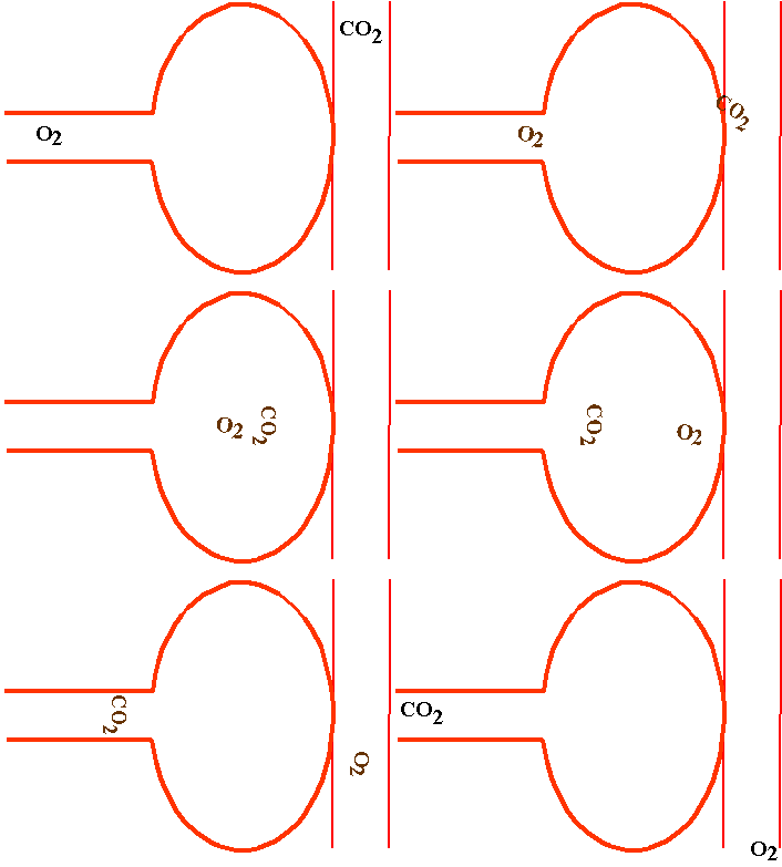
Alveolar Ventilation
I. The Standard Lung Volumes and Capacities - depend on the mechanics of the lungs and chest wall, and on respiratory muscle activity. Obtained under specific conditions. Must be normalized for subject s height, weight, age, sex, etc. so they are compared to data from a table of predicted values (Levitzky Fig 3-1). The standard lung volumes and capacities can change with body position (Levitzky Fig 3-2) and in pathologic states (Levitzky Fig 3-3).
A. Total Lung Capacity (TLC) - the volume of air in the lungs after a maximal inspiratory effort. Determined by the strength of contraction of the inspiratory muscles in opposition to the inward elastic recoil of the lungs and chest wall. About 6 liters in a healthy 70 kg adult.
B. Residual Volume (RV) - the volume of gas left in the lungs after a maximal forced expiration. Determined by the force generated by the muscles of expiration and the inward elastic recoil of the lungs as they oppose the outward elastic recoil of the chest wall. Dynamic compression of the airways during the forced expiratory effort is also an important determinant of the residual volume - as airway collapse occurs gas is trapped in the alveoli. The residual volume of a healthy 70-kg adult is 1.5 liters.
C. Functional Residual Capacity (FRC) - the volume of gas remaining in the lungs at the end of a normal tidal expiration. Balance point between the inward elastic recoil of the lungs and the outward elastic recoil of the chest wall. About 3 liters in a healthy 70-kg adult.
D. Tidal Volume (VT) - the volume of air entering or leaving the nose or mouth per breath. During normal, quiet breathing (eupnea) the tidal volume of a 70-kg adult is about 500 ml per breath.
E. Expiratory Reserve Volume (ERV) - the volume of gas expelled from the lungs during a maximal forced expiration that starts at the end of normal tidal expiration. About 1.5 liters.
F. Inspiratory Reserve Volume (IRV) - the volume of gas inhaled into the lungs during a maximal forced inspiration starting at the end of a normal tidal inspiration. About 2.5 liters.
G. Inspiratory Capacity (IC) - the volume of air inhaled into the lungs during a maximal inspiratory effort that begins at the end of a normal tidal expiration. About 3 liters.
H. Vital Capacity (VC) - the volume of air expelled from the lungs during a maximal forced expiration starting after a maximal forced inspiration. About 4.5 liters.
II. Measurement of the lung volumes and capacities
A. Spirometry - must be able to exchange the volume to be determined with the spirometer. Therefore cannot use spirometry to determine the RV, or the FRC and TLC, which contain the RV (Levitzky Fig 3-4).
B. Determination of the FRC, RV, and TLC
1. Nitrogen washout technique
2. Helium dilution technique (Levitzky Fig 3-5).
3. Body plethysmograph technique - includes trapped gas (Levitzky Fig 3-6).
III. Alveolar ventilation and dead space A. Alveolar ventilation ( A) is defined as the volume of air entering and leaving the alveoli per minute. Air ventilating the anatomic dead space (VD) (Levitzky Fig 3-7), where no gas exchange occurs, is not included:
VT = VD + VA
VA = VT - VD
n(VA) = n(VT) - n(VD)
A = E - D
IV. Determination of dead space
A. Anatomic dead space (VD)
1. Approximately 1 ml/lb body weight in normal adult. Neural reflexes, traction or compression and pathologic changes can alter the anatomic dead space.
2. Fowler's method - monitor expired [N2] after single breath of O2 (Levitzky Fig 3-8).
B. Physiologic dead space
1. Physiologic dead space = anatomic dead space plus alveolar dead space.
2. Alveolar dead space - alveoli that are ventilated but not perfused. There is normally no alveolar dead space, so physiologic dead space equals anatomic dead space.
3. Determined by using the Bohr equation:
4. Arterial PCO2 is normally equal to alveolar (end-tidal) PCO2 When arterial PCO2 > alveolar PCO2, then alveolar dead space is present and physiologic dead space > anatomic dead space (Levitzky Fig 3-9).
V. The effects of alveolar ventilation on alveolar PCO2 and PO2:
A. PACO2 If alveolar ventilation is doubled (and carbon dioxide production is unchanged), then the alveolar and arterial PCO2 are reduced by one-half. If alveolar ventilation is cut in half, near 40 mm Hg, then alveolar and arterial PCO2 will double (Levitzky Fig 3-10 top).
B. PO2 - As alveolar ventilation increases, the alveolar PO2 also increases. Doubling alveolar ventilation cannot double alveolar PO2 in a person whose alveolar PO2 is already 104 mm Hg because the highest PAO2 one can achieve (breathing air at sea level) is the inspired PO2 of about 149 mm Hg (Levitzky Fig 3-10 bottom). The alveolar PO2 can be calculated by using the alveolar air equation:
PAO2 = PIO2 - + F
C. PIO2 = inspired PO2 = FIO2 (Pbarom - PH2O);
PH2O is almost always 47 torr by the end of the airway.
R = respiratory exchange ratio = F = a small correction factor
VI. Regional distribution of alveolar ventilation
A. More ventilation of lower (with respect to gravity) regions of the lung than upper regions of the lung when breathe around the FRC (Levitzky Fig 3-11).
B. Reason: Intrapleural ( intrathoracic ) pressure is less negative at the bottom of the lung than at the top because of the weight of the lung and the configuration of the chest wall. There is a gradient of intrapleural pressure that increases 0.2 - 0.3 cm H2O per cm vertical displacement as you move down the pleural surface.
C. Therefore the transpulmonary pressure gradient is greater at the top of the lung than at the bottom.
D. Therefore the alveoli at the top of the lung are at a higher, less compliant point on the pressure-volume curve than those at the bottom (Levitzky Fig 3-12).
E. Therefore the alveoli at the bottom of the lung increase their volume more with each inspiration and decrease their volume more with each expiration during eupnea (from FRC).
F. Changes in this regional distribution of ventilation can occur at different lung volumes. For example, near the Residual Volume most ventilation occurs at upper alveoli(Levitzky Fig 3-13).
VII. Regional distribution of lung volume.
A. FRC and RV mainly in upper regions
B. IRV mainly in lower regions
VIII. Closing volume and closing capacity (Levitzky Fig 3-14).
A. Airway closure should first occur in lower regions ( dependent regions ) of the lung:
1. Intrapleural pressure is more positive in lower regions
2. Alveoli are smaller - less alveolar elastic recoil so less traction holding small airways open.
B. In emphysema and in old age airways in lower regions of the lung may be closed at the FRC. Thus the closing capacity > FRC.
IX. The effects of aging on the respiratory system (Levitzky Fig 3-15).
A. Decreased alveolar elastic recoil
B. Chest wall less compliant
C. Less strength of respiratory muscles
D. Loss of alveolar surface area, decreased diffusing capacity
E. More mismatch, lower PaO2.
Copyright 2000 M. G. LEVITZKY
Last updated Monday July 15, 2013 3:15 PM
LSUHSC is an equal opportunity educator and employer.
The statements found on this page are for informational purposes only. While every effort is made to ensure that this information is up-to-date and accurate, for official information please consult a printed University publication.
The views and opinions expressed in this page are strictly those of the page author. The contents of this page are not reviewed or approved by LSUHSC.
This page is maintained by webmaster-arc: mgiaim@lsuhsc.edu
