Case of the Month - March 2024
Michael Webber, DO, Haibo Wang, MD, Wenjing Qiu, MD, Tracy Dewenter, MD
A 48-year-old female presented to her primary care physician for evaluation of kidney dysfunction that began recently. A computed tomography scan showed a 3.3 cm enhancing, endophytic mass in the right lower pole of her kidney. The patient underwent a right radical nephrectomy. A gross pathology examination revealed a red-tan, soft, well-circumscribed mass (4.4 x 3.8 x 3.6 cm) with areas of hemorrhage and necrosis (~50%), in the lower pole of the kidney. Representative hematoxylin and eosin (H&E)-stained sections at low, medium, and high power and some immunohistochemical stains are demonstrated in the figures below.
Figure 1.
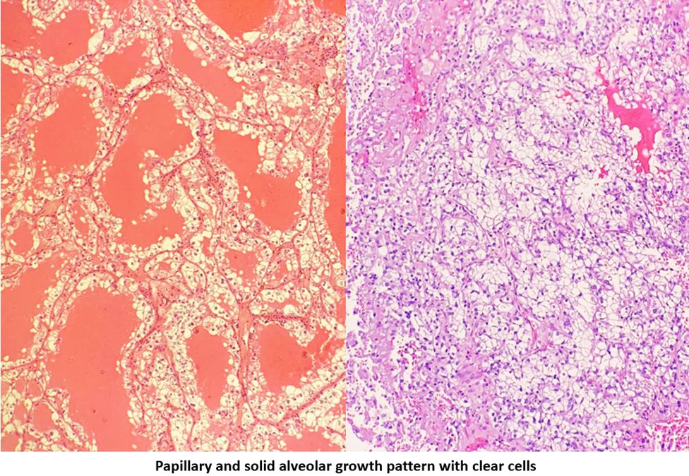
Figure 2.
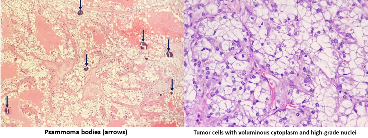
Figure 3.
Immunohistochemical (IHC) stains show positive expression of AMACR and CD10, as well as focal positivity for CA IX. There was no significant expression of CK7, CD117, CK-HMW, or GATA-3 within the tumoral cells.

What is the most likely diagnosis?
- Chromophobe Renal Cell Carcinoma (RCC)
- Clear cell RCC
- Papillary RCC
- Translocation RCC
- Clear cell papillary renal cell tumor