February 2025 - Case of the Month
Angelle Jolly, MD, Darshan Trivedi, MD, and Ellen Connor, MD, PhD
An 81-year-old female with a history of metastatic nasal cavity cancer, hypertension, failure to thrive, and chronic obstructive pulmonary disease (COPD) presented with acute onset of shortness of breath and hypoxia. Imaging upon arrival was significant for segmental and subsegmental pulmonary emboli with multiple bilateral pulmonary nodules, which were attributed to metastatic involvement of the patient’s nasal cavity cancer. Anticoagulation therapy was initiated, and she developed recurrent epistaxis and dark stools. Shortly after, she became unresponsive and developed agonal breathing. She was transitioned to comfort care measures and pronounced dead. An unrestricted autopsy was requested by the decedent’s next of kin.
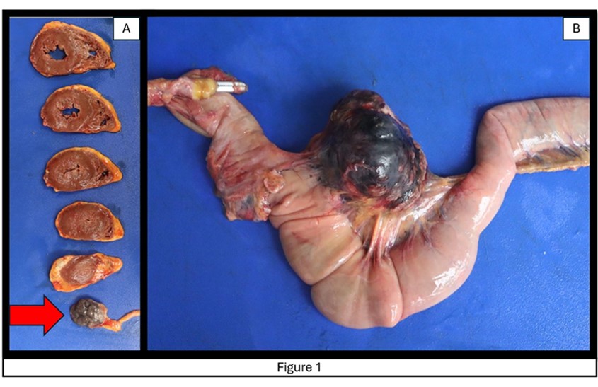
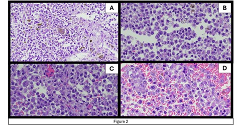
At autopsy, multiple lesions were noted in the cardiac apex, the mesentery (Figure 1), the left lung, the left thyroid lobe, the peripancreatic space, and the right cerebral caudate. The lesions in the cardiac apex, the mesentery, the peripancreatic space, the left thyroid lobe, and the right cerebral caudate were all well-circumscribed, nodular, and black. The lesion in the left lung was well-defined, white-tan, and firm. Sections were taken for microscopic examination (Figure 2) from the cardiac apex (A), mesenteric nodule (B), pancreas (C), and thyroid (D).


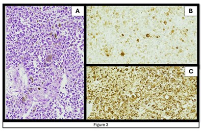
Slides of the antemortem nasal cavity lesion biopsy revealed epithelioid pleomorphic cells with prominent nucleoli, similar to the cells identified in the cardiac apex, mesentery, peripancreatic space, and thyroid. HMB-45 and Melan-A immunostains were performed and were positive.
The cardiac apex lesion (Figure 3) was stained with Melan-A (B) and HMB-45 (C).

Question: Which of the following, if present, is most helpful in distinguishing this entity from cutaneous melanoma?
- HMB-45 positivity
- NRAS mutation
- History of UV exposure
- KIT mutation
- History of radiation exposure
Click here for answer and discussion
